Hyaline Cartilage Drawing, It contains no nerves or blood vessels, and its structure is relatively simple.
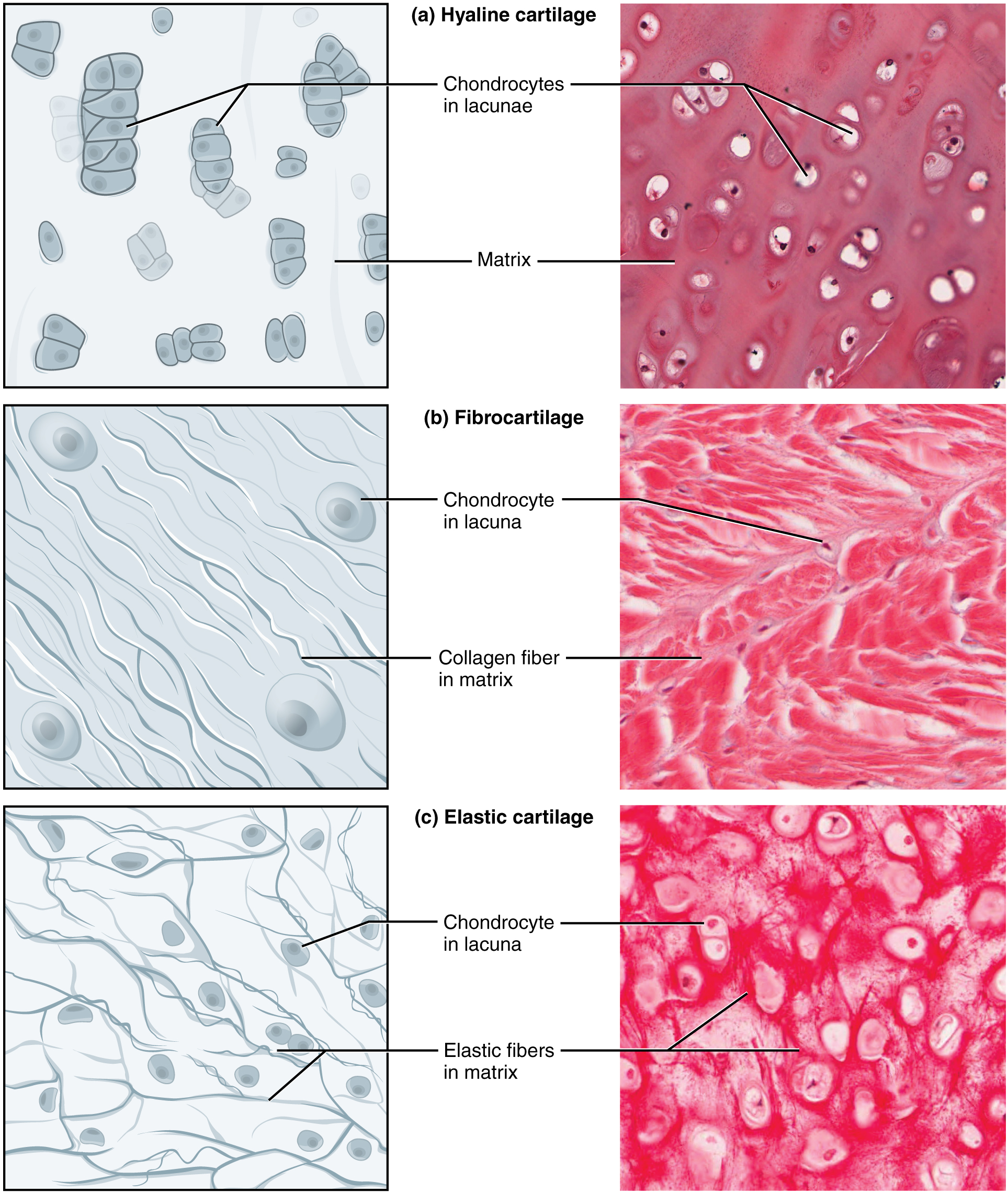
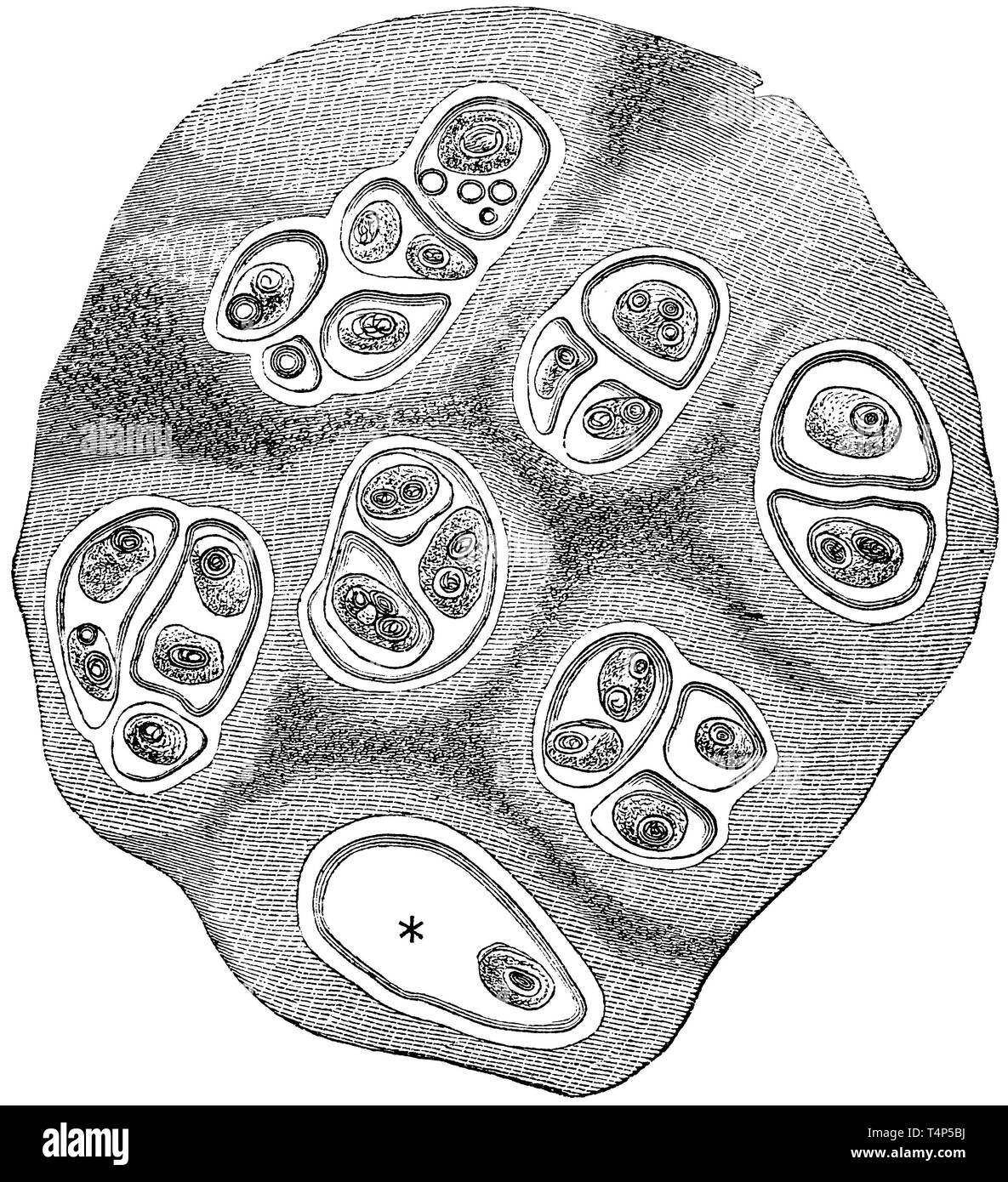
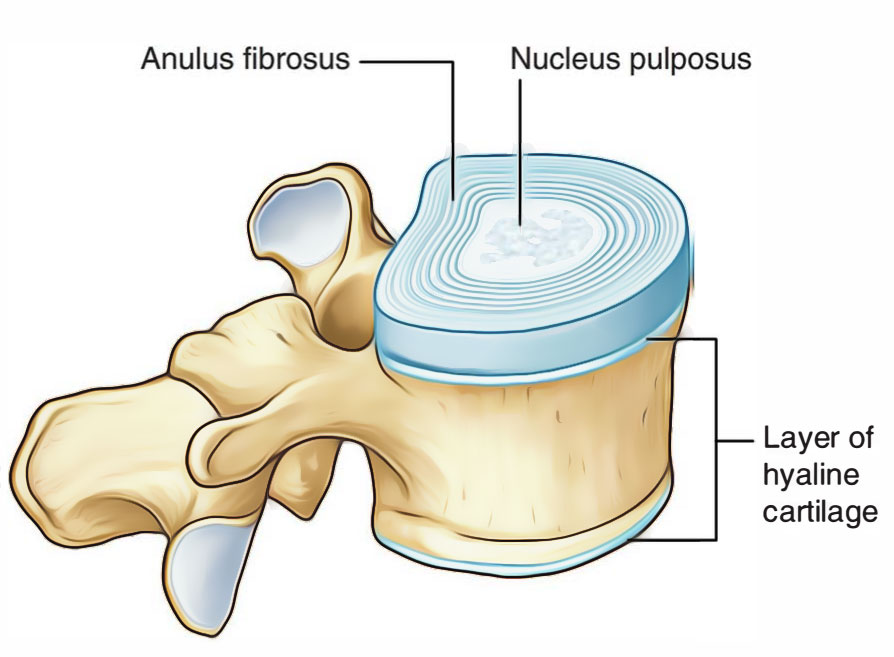
Hyaline Cartilage Drawing - Use the image slider below to learn how to use a microscope to identify and study hyaline cartilage on a microscope slide of the trachea. Now you will able to identify the hyaline cartilage histology slide under a light microscope with proper identification points. #biology #hyalinecartilage animaltissue #class11 #hscbiology #maharashtrastateboard2021 #biology2021 #biologydiagrams #icse #cbse this diagram shows how to draw hyaline. Isogenous groups and interstitial growth results when chondrocytes divide and produce extracellular matrix. There are several oval pieces of hyaline cartilage on this slide. It is also most commonly found in the ribs, nose, larynx, and trachea. It has a unique structure that is organised into specific zones. This image shows a cross section of a cartilage ring that supports the trachea and maintains the openness (patency) of the airway. Web articular cartilage is a type of hyaline cartilage that is found on the surface of bones in synovial joints. Comparison with standard mr imaging and correlation with arthroscopy. Web image 2 of 19. Isogenous groups and interstitial growth results when chondrocytes divide and produce extracellular matrix. This article will focus on important features of hyaline cartilage, namely its matrix, chondrocytes, and perichondrium. Web hyaline cartilage, the most abundant type of cartilage, plays a supportive role and assists in movement. There are several oval pieces of hyaline cartilage on. The word hyaline is derived from the greek word ‘ hyalos ’, which means ‘ glassy ’. Formed by the process of chondrogenesis, the resulting chondrocytes are capable of producing large amounts of collagenous extracellular matrix and ground substance, which together form cartilage itself. Comparison with standard mr imaging and correlation with arthroscopy. Step by step drawing of histology of. Web type i collagen (col i) and hyaluronic acid (ha), derived from the extracellular matrix (ecm), have found widespread application in cartilage tissue engineering. A joint of the jaw that connects it to the temporal bones of the skull. Territorial matrix lies immediately around each isogenous group and is high in glycosaminoglycans. Web hyaline cartilage, the most common type of. It is also most commonly found in the ribs, nose, larynx, and trachea. Web type i collagen (col i) and hyaluronic acid (ha), derived from the extracellular matrix (ecm), have found widespread application in cartilage tissue engineering. Unlike most hyaline cartilage, articular hyaline cartilage does not have a. Web articular cartilage is a type of hyaline cartilage that is found. Web this is the best guide to learn hyaline cartilage histology with labeled diagram and real slide pictures. Comparison with standard mr imaging and correlation with arthroscopy. This image shows a cross section of a cartilage ring that supports the trachea and maintains the openness (patency) of the airway. Web hyaline cartilage is the most prevalent type, forming articular cartilages. It is also most commonly found in the ribs, nose, larynx, and trachea. Formed by the process of chondrogenesis, the resulting chondrocytes are capable of producing large amounts of collagenous extracellular matrix and ground substance, which together form cartilage itself. Use the image slider below to learn how to use a microscope to identify and study hyaline cartilage on a. Comparison with standard mr imaging and correlation with arthroscopy. Now you will able to identify the hyaline cartilage histology slide under a light microscope with proper identification points. Use the image slider below to learn more about the characteristics of hyaline cartilage. #biology #hyalinecartilage animaltissue #class11 #hscbiology #maharashtrastateboard2021 #biology2021 #biologydiagrams #icse #cbse this diagram shows how to draw hyaline. Web. Web be able to recognize and differentiate the three major cartilage types (hyaline, elastic and fibrocartilage) in light microscope images and know how they differ in composition and examples where each type is found in the human body. #biology #hyalinecartilage animaltissue #class11 #hscbiology #maharashtrastateboard2021 #biology2021 #biologydiagrams #icse #cbse this diagram shows how to draw hyaline. It is also most commonly. Web hyaline cartilage, the most common type of cartilage, is composed of type ii collagen and chondromucoprotein and often has a glassy appearance. Note the numerous chondrocytes in this image, each located within lacunae and surrounded by. A joint of the jaw that connects it to the temporal bones of the skull. Web during embryonic development, hyaline cartilage serves as. Note the numerous chondrocytes in this image, each located within lacunae and surrounded by. Formed by the process of chondrogenesis, the resulting chondrocytes are capable of producing large amounts of collagenous extracellular matrix and ground substance, which together form cartilage itself. Territorial matrix lies immediately around each isogenous group and is high in glycosaminoglycans. The word hyaline is derived from. It has a unique structure that is organised into specific zones. This image shows a cross section of a cartilage ring that supports the trachea and maintains the openness (patency) of the airway. Use the image slider below to learn how to use a microscope to identify and study hyaline cartilage on a microscope slide of the trachea. Web hyaline cartilage is a type of connective tissue found in areas such as the nose, ears, and trachea of the human body. Web draw it easy!!! A joint of the jaw that connects it to the temporal bones of the skull. Use the image slider below to learn more about the characteristics of hyaline cartilage. Cells that form and maintain the cartilage. Examine them with low power first. A type of cartilage found on many joint surfaces; Web in adults, hyaline cartilage is located in the articular surfaces of movable joints, in the walls of the respiratory tracts (nose, larynx, trachea, and bronchi), in the costal cartilages, and. Formed by the process of chondrogenesis, the resulting chondrocytes are capable of producing large amounts of collagenous extracellular matrix and ground substance, which together form cartilage itself. #biology #hyalinecartilage animaltissue #class11 #hscbiology #maharashtrastateboard2021 #biology2021 #biologydiagrams #icse #cbse this diagram shows how to draw hyaline. 8.1k views 4 years ago. The word hyaline is derived from the greek word ‘ hyalos ’, which means ‘ glassy ’. Web be able to recognize and differentiate the three major cartilage types (hyaline, elastic and fibrocartilage) in light microscope images and know how they differ in composition and examples where each type is found in the human body.
Connective Tissue Supports and Protects · Anatomy and Physiology

Hyaline Cartilage Drawing YouTube

Illustrations Hyaline Cartilage General Histology

Hyaline cartilage structure and biochemical composition. Schematic

How to draw histology of hyaline cartilage ? YouTube

How to Draw Hyaline Cartilage Simple and easy steps Biology Exam

Hyaline Cartilage Drawing

Hyaline Cartilage Labeled Diagram

Hyaline Cartilage Drawing

Hyaline Cartilage Drawing
Now You Will Able To Identify The Hyaline Cartilage Histology Slide Under A Light Microscope With Proper Identification Points.
Each Piece Of Cartilage Is Surrounded By A Perichondrium.
Unlike Most Hyaline Cartilage, Articular Hyaline Cartilage Does Not Have A.
Web Image 2 Of 19.
Related Post: