Drawing Of Cardiac Muscle, Web cardiac muscle tissue is one of the three types of muscle tissue in your body.
Drawing Of Cardiac Muscle - Web cardiac muscle tissue is a specialized, organized type of tissue that only exists in the heart. Web this article describes the characteristics, components and function of the cardiac muscle tissue, including clinical points. Highly coordinated contractions of cardiac muscle pump blood into the vessels of the circulatory system. These are striated and involuntary muscles that are supplied by autonomic nerve fibres. Highly coordinated contractions of cardiac muscle pump blood into the vessels of the circulatory system. Web the cardiac muscle under a microscope shows a short cylindrical fiber with a centrally placed oval nucleus. Web review the cardiac muscle cells which make up the myocardium portion of the heart wall in this interactive tutorial, and test yourself in the quiz. The middle layer, composed of muscle tissue that enables heart contractions. Web cardiac muscle (or myocardium) makes up the thick middle layer of the heart. It is one of three types of muscle in the body, along with skeletal and smooth. Web for instance, the risk of major complications during lateral trans iliac (lti) si joint fusion (cpt code 27279) is lower than the risks associated with other obl. It is responsible for keeping the heart pumping and blood circulating. Web cardiac muscle tissue is only found in the heart. You will find some unique features in cardiac muscle. The video. Web cardiac muscle differs from skeletal muscle in that it exhibits rhythmic contractions and is not under voluntary control. Web for instance, the risk of major complications during lateral trans iliac (lti) si joint fusion (cpt code 27279) is lower than the risks associated with other obl. The outermost layer protects the heart and reduces. Web this article describes the. It is one of three types of muscle in the body, along with skeletal and smooth. The other two types are skeletal muscle tissue and smooth muscle tissue. The rhythmic contraction of cardiac. The middle layer, composed of muscle tissue that enables heart contractions. The outermost layer protects the heart and reduces. Web review the cardiac muscle cells which make up the myocardium portion of the heart wall in this interactive tutorial, and test yourself in the quiz. It is responsible for keeping the heart pumping and blood circulating. The outermost layer protects the heart and reduces. Highly coordinated contractions of cardiac muscle pump blood into the vessels of the circulatory system.. The other two types are skeletal muscle tissue and smooth muscle tissue. It is one of three types of muscle in the body, along with skeletal and smooth. Highly coordinated contractions of cardiac muscle pump blood into the vessels of the circulatory system. These inner and outer layers of. Web the cardiac muscle or the myocardium forms the musculature of. Web for instance, the risk of major complications during lateral trans iliac (lti) si joint fusion (cpt code 27279) is lower than the risks associated with other obl. Web the cardiac muscle or the myocardium forms the musculature of the heart. Identify and describe the components of the conducting system that distributes electrical impulses through the heart; The rhythmic contraction. These are striated and involuntary muscles that are supplied by autonomic nerve fibres. Highly coordinated contractions of cardiac muscle pump blood into the vessels of the circulatory system. Web for instance, the risk of major complications during lateral trans iliac (lti) si joint fusion (cpt code 27279) is lower than the risks associated with other obl. Web cardiac muscle, also. Highly coordinated contractions of cardiac muscle pump blood into the vessels of the circulatory system. The myocardium is the muscular middle layer of the heart wall that contains the cardiac muscle tissue. The outermost layer protects the heart and reduces. Highly coordinated contractions of cardiac muscle pump blood into the vessels of the circulatory system. The middle layer, composed of. Identify and describe the components of the conducting system that distributes electrical impulses through the heart; The other two types are skeletal muscle tissue and smooth muscle tissue. Learn this topic now at kenhub! It is one of three types of muscle in the body, along with skeletal and smooth. Web review the cardiac muscle cells which make up the. Web cardiac muscle tissue is a specialized, organized type of tissue that only exists in the heart. You will find some unique features in cardiac muscle. Learn this topic now at kenhub! Web cardiac muscle, also known as heart muscle, is the layer of muscle tissue which lies between the endocardium and epicardium. It is one of three types of. The myocardium is the muscular middle layer of the heart wall that contains the cardiac muscle tissue. The rhythmic contraction of cardiac. Identify and describe the components of the conducting system that distributes electrical impulses through the heart; Web this article describes the characteristics, components and function of the cardiac muscle tissue, including clinical points. You will find some unique features in cardiac muscle. Web cardiac muscle tissue is only found in the heart. Web cardiac muscle tissue is only found in the heart. Learn this topic now at kenhub! Web cardiac muscle tissue is a specialized, organized type of tissue that only exists in the heart. Web review the cardiac muscle cells which make up the myocardium portion of the heart wall in this interactive tutorial, and test yourself in the quiz. Web for instance, the risk of major complications during lateral trans iliac (lti) si joint fusion (cpt code 27279) is lower than the risks associated with other obl. These are striated and involuntary muscles that are supplied by autonomic nerve fibres. Web describe the structure of cardiac muscle; These inner and outer layers of. Web cardiac muscle, also known as heart muscle, is the layer of muscle tissue which lies between the endocardium and epicardium. It is responsible for keeping the heart pumping and blood circulating.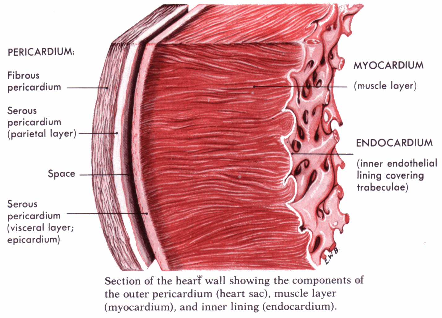
Which is the cardiac muscle layer of the heart? Socratic
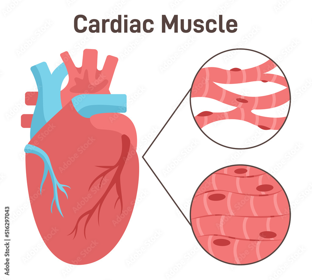
Cardiac muscle fibers' structure. Heart muscle tissue, anatomy Stock

Example of a cardiac muscle
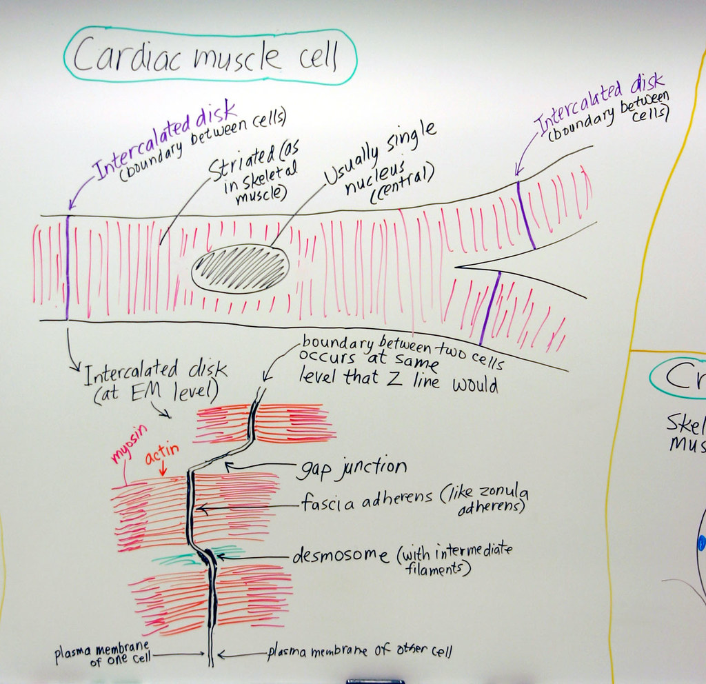
Muscle Cardiac Muscle Cell A hand drawn sketch by Dr. Chr… Flickr
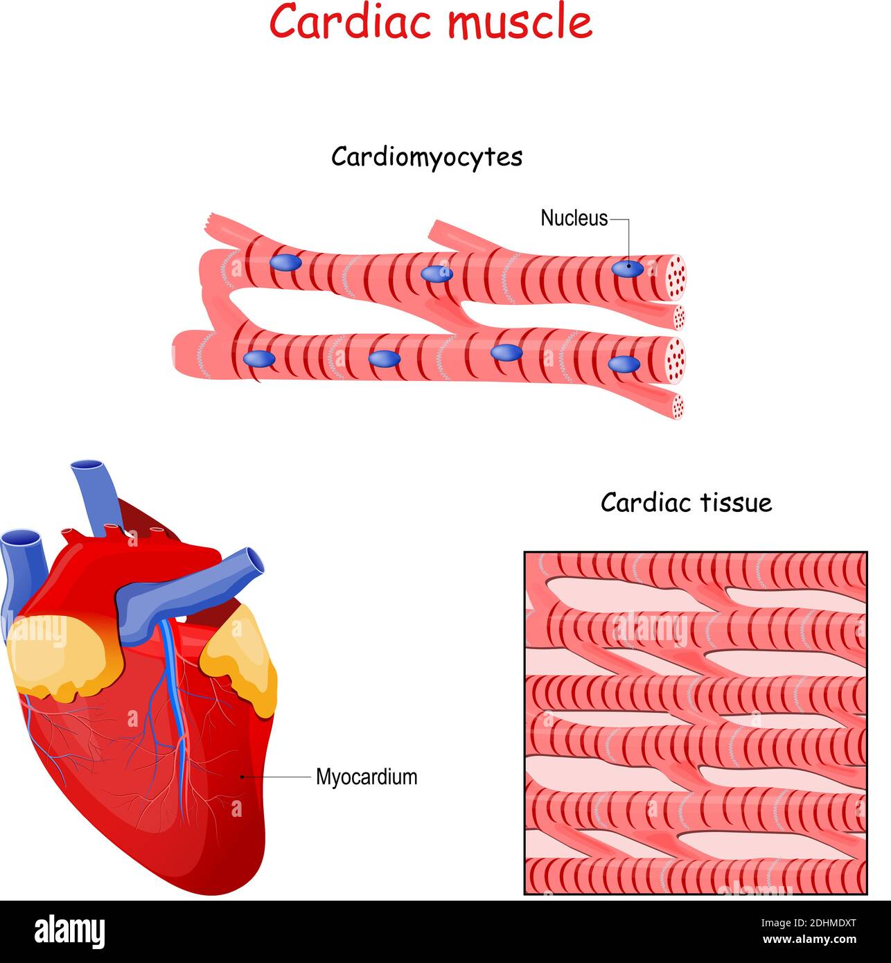
Cardiac Muscle Structure

How to draw " Cardiac Muscles" step by step in a very easy way Type
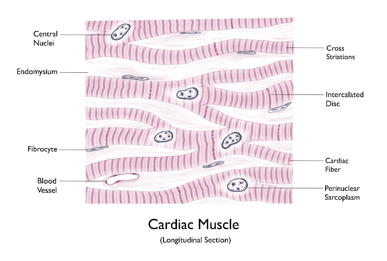
Cardiac Muscle (longitudinal section)

Stockvector Heart anatomy colored sketch. Anatomic human cardiac muscle
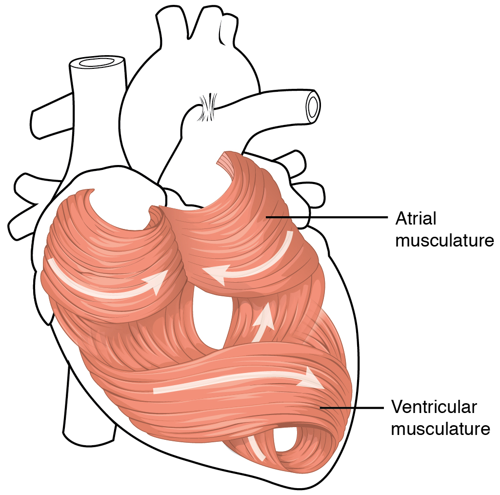
Heart Anatomy · Anatomy and Physiology

Cardiac Muscle Vector Illustration Diagram, Anatomical Scheme with
The Middle Layer, Composed Of Muscle Tissue That Enables Heart Contractions.
The Video Describes The Summary Of The Whole Topic Of The Muscle.
It Is One Of Three Types Of Muscle In The Body, Along With Skeletal And Smooth.
Myocardium Makes Up The Majority Of The Thickness And Mass.
Related Post: